
The Alarm in the Corner of the Eye: An Immediate Cold Open on Sudden Tissue Shifts
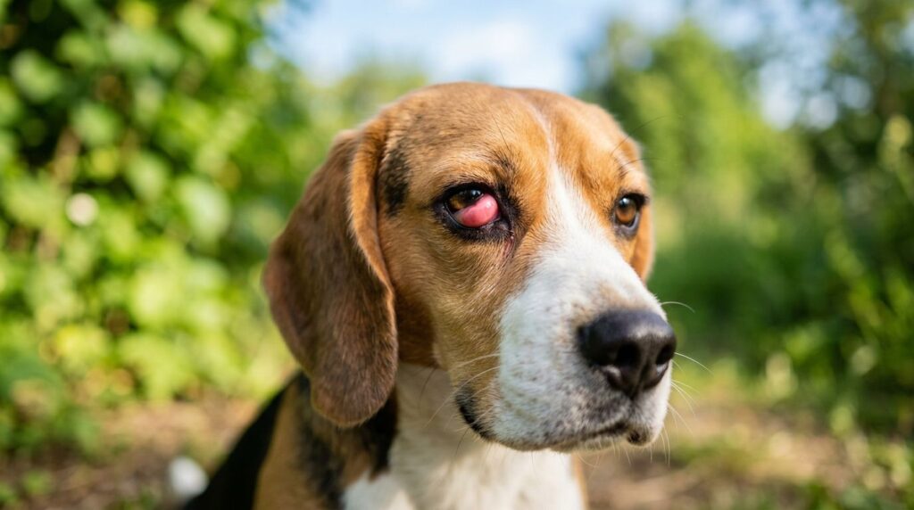
A smooth, fleshy, bright pink mass can appear almost instantly in the inner corner of a dog’s eye, catching pet owners completely by surprise during a routine morning greeting or an evening play session. This sudden structural shift looks undeniably shocking. Many families experience an immediate wave of panic when they first see the tissue, naturally assuming that their companion has developed an aggressive, fast-growing tumor or a dangerous, deep-seated ocular infection. The dramatic visual appearance of the red nodule often drives a frantic search for answers, as the tissue can swell to a size that partially obscures the animal’s vision.
This distinct ocular anomaly is known clinically as a prolapse of the nictitating membrane gland, a condition commonly referred to as canine cherry eye. While the sudden protrusion appears to be a disfiguring medical emergency, it is actually a benign, purely structural failure of the tissues supporting the eye components. It is a highly treatable condition that responds exceptionally well to modern veterinary care. Resolving the issue successfully requires a calm approach and professional surgical correction rather than panic, delayed monitoring, or risky experimentation with unverified home remedies.
Table of Contents
- The Hidden Anchor: Anatomical Failure of the Third Eyelid Gland
- The Danger of Delay: How Procrastination Spells Secondary Optical Damage
- The Surgical Remedy over Mass Excision: Preserving Vital Moisture Chambers
- Advanced Ophthalmology and Wellness on SW Nimbus Ave: Our Tactical Defense Framework
- Local, Unbiased Pet Care: Why Independent Care Paths Protect Tigard Households Better
- Washington County Ocular Reference: Pressing Questions for Local Dog Owners
- Reclaiming Your Dog’s Optical Health and Visual Comfort
The Hidden Anchor: Anatomical Failure of the Third Eyelid Gland
Understanding why this tissue shift occurs requires a brief look at the unique anatomy of the canine eye. Unlike humans, dogs possess three distinct eyelids. The third eyelid, technically termed the nictitating membrane, sits tucked away out of sight in the inner corner of the eye socket, near the tear duct. This specialized membrane functions as an extra protective shield, sweeping horizontally across the clear surface of the eye during blinking to clear away microscopic dust, pollen, and environmental debris.
Deep within the base of this third eyelid rests a vital, pink gland that is responsible for producing roughly thirty to fifty percent of the watery component of the dog’s natural tear film. Under normal conditions, this tear gland stays completely hidden from view, anchored securely to the lower inner rim of the orbital bone by a small, flexible band of fibrous connective tissue. This connective tissue anchor acts as a tiny bungee cord, holding the gland down in its proper anatomical pocket while allowing the third eyelid to move freely.
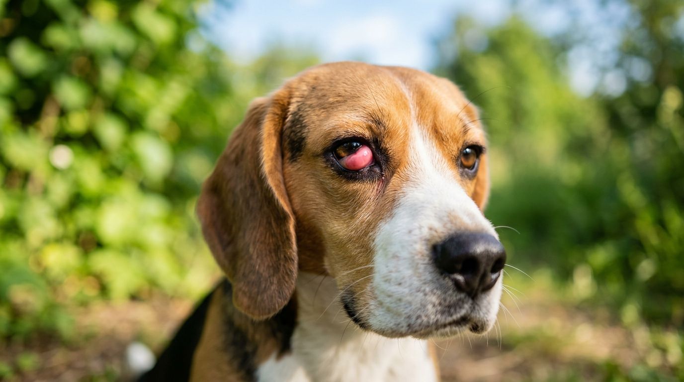
When this fibrous anchor is genetically weak, stretches out over time, or suffers a microscopic tear during vigorous play, the structural integrity of the pocket is lost. With the anchor gone, the natural pressure within the eye socket forces the tear gland to flip upward and pop completely out of its hidden home. Once the sensitive glandular tissue is exposed to the open air, the delicate mucous membranes quickly become inflamed. The gland swells into the signature round, red, cherry-like mass that gives the condition its common name.
- Genetics play a foundational role in this structural failure, with specific breeds common across Washington County showing a strong predisposition, including English Bulldogs, French Bulldogs, Cocker Spaniels, Beagles, Boston Terriers, and Mastiffs.
- The issue frequently presents in young puppies under one year of age, a developmental window when the connective tissue anchors are still growing and may fail to support the rapidly changing structures of the skull.
- The protruding pink mass is an inflamed, displaced internal gland rather than a foreign tumor, a cancerous growth, or a contagious infection, meaning it cannot spread to other pets in the household.
The Danger of Delay: How Procrastination Spells Secondary Optical Damage
Leaving a prolapsed third eyelid gland untreated out of a hope that it will simply slide back into place on its own poses a serious risk to an animal’s long-term eye health. The longer the sensitive glandular tissue remains displaced and exposed to the elements, the more its physical condition deteriorates. The delicate membranes are not designed to withstand constant exposure to dry household air, windy Pacific Northwest weather, or airborne dust particles. As the exposed gland dries out, the swelling intensifies, restricting local blood circulation and causing the tissue to become increasingly raw, irritated, and chronically inflamed.
This persistent physical discomfort creates a constant, nagging sensation akin to having a large piece of grit trapped permanently beneath the eyelid. Dogs dealing with this chronic irritation will naturally look for ways to relieve the itch, frequently rubbing their faces forcefully against carpets, couches, or rough tree bark during walks. They may also paw frantically at their eyes with their front dewclaws. This self-inflicted trauma introduces environmental bacteria into the eye socket and creates a substantial risk of serious, permanent physical injury to the surrounding ocular structures.
- Progressive reduction in essential tear film production occurs as the exposed, inflamed gland becomes chronically damaged, leading directly to a painful condition known as chronic dry eye, or keratoconjunctivitis sicca.
- Continuous exposure to environmental debris triggers painful secondary bacterial conjunctivitis, which fills the eye socket with a thick, yellow or green mucus discharge that crusts along the eyelids.
- Corneal scratching and the development of deep, slow-healing ocular ulcers can manifest rapidly from the friction of the protruding mass or from the dog’s persistent pawing at their face.
Once chronic dry eye develops due to long-term gland damage, the eye loses its primary defense mechanism against infection and friction. The cornea can become severely scarred, dry, and pigmented, a progressive change that can eventually lead to permanent blindness. Seeking prompt veterinary intervention before these secondary complications take hold is essential to preserve your pet’s natural vision and prevent a straightforward structural fix from turning into a complex, lifelong medical problem.
The Surgical Remedy over Mass Excision: Preserving Vital Moisture Chambers
In the past, the standard veterinary approach to resolving cherry eye was a fast, cosmetic procedure that involved simply clipping the protruding red bump off at the base. While this method instantly removed the unsightly mass and restored the normal appearance of the eye, long-term clinical data eventually revealed disastrous biological consequences. Completely removing the gland solves the cosmetic problem but permanently robs the eye of up to half of its natural moisture-producing capacity. Dogs who underwent this mass excision almost inevitably developed severe, irreversible dry eye later in life, forcing their owners to administer expensive artificial tear drops multiple times a day for the rest of the animal’s existence.
Modern veterinary surgical excellence completely rejects the practice of removing healthy, functional glandular tissue. Today, the golden standard of canine soft-tissue surgery focuses entirely on preservation, restoration, and anatomical realignment. Advanced surgical procedures are designed to create a secure new tissue pocket or anchor the displaced gland back down into its natural home beneath the third eyelid, protecting its vital role in tear production.
The most common modern technique is known as the pocket method, or the Morgan Pocket technique. During this delicate procedure, a skilled veterinary surgeon makes small, precise incisions in the mucous membrane surrounding the exposed gland, creating a secure, hidden structural envelope. The living gland is then gently tucked back down into this custom pocket, and the edges of the tissue are closed over it using incredibly fine, dissolving suture material. The knots are carefully placed on the outside surface of the eyelid to ensure they never rub against or scratch the delicate cornea. This sophisticated approach preserves the moisture-producing cells completely, allowing the gland to heal fully and continue lubricating the eye for the rest of your dog’s life.
Advanced Ophthalmology and Wellness on SW Nimbus Ave: Our Tactical Defense Framework
Nimbus Pet Hospital approaches ocular conditions with a combination of cutting-edge diagnostic technology, modern surgical instrumentation, and deeply compassionate handling. Located on SW Nimbus Ave, our clinic serves as a comprehensive medical resource for families navigating sudden health changes in their pets. We understand that eye issues can be highly stressful for animals, which is why we structure our evaluations to minimize anxiety and deliver rapid, accurate results under one roof.
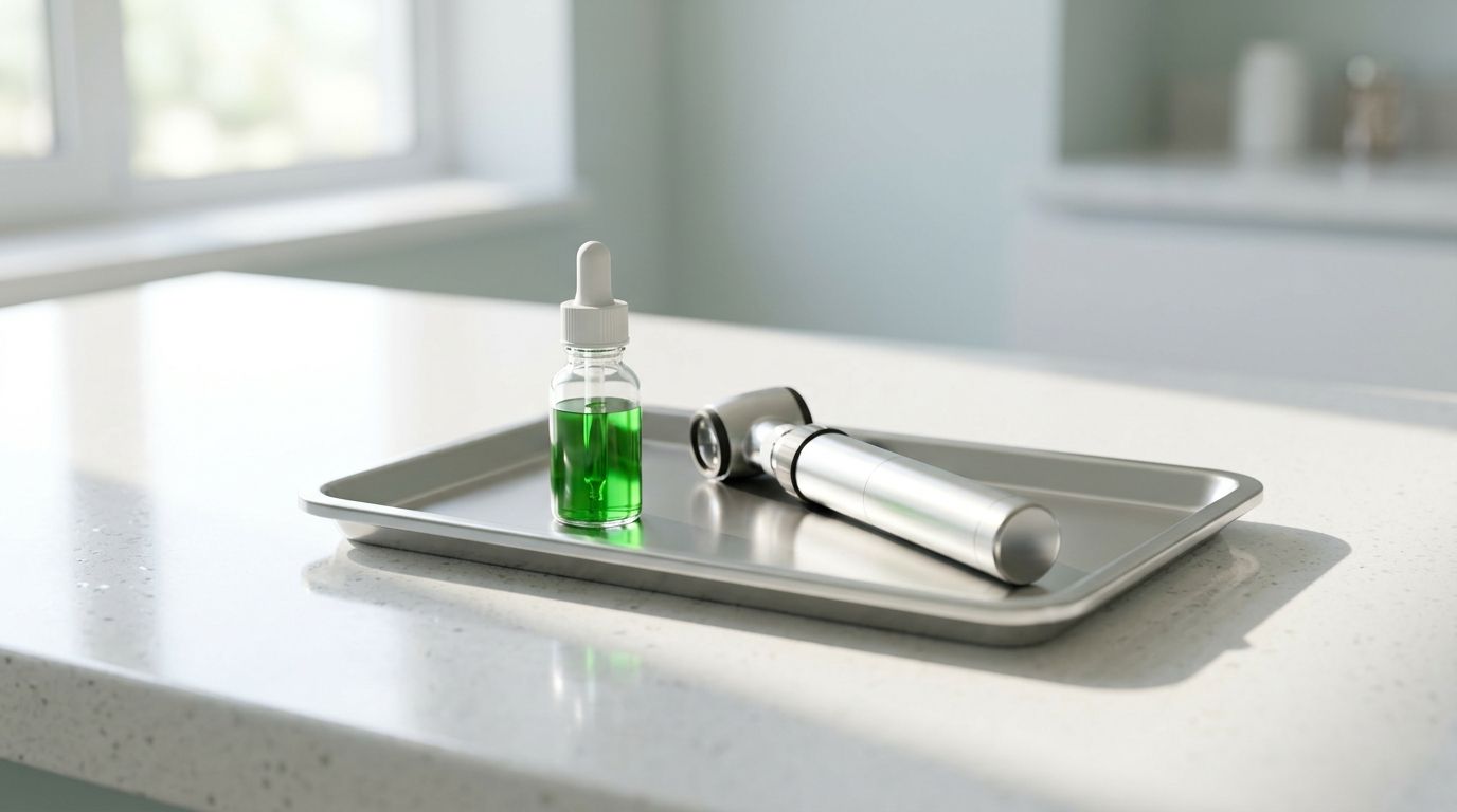
Our comprehensive wellness examinations include detailed eye assessments whenever an owner notices a change in their pet’s ocular appearance. Before suggesting a surgical plan, our veterinary team performs precise diagnostic checks to evaluate the overall health of the eye. We use Schirmer tear tests to measure baseline tear film production, ensuring we understand the gland’s current functional capacity. Additionally, we apply specialized fluorescein staining dyes to the surface of the eye, using a specialized light source to screen for any hidden corneal ulcers or micro-scratches caused by the prolapsed tissue.
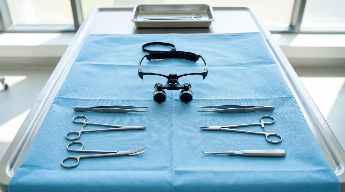
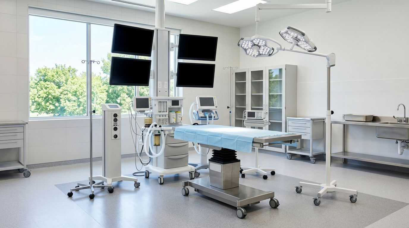
When surgery is required, our advanced soft-tissue surgical suites provide a highly secure environment for delicate facial procedures. We utilize high-magnification surgical loupes and micro-instrumentation to perform pocket repairs with exceptional precision, ensuring every suture is perfectly placed. Patient safety is our absolute priority, which is why our facility uses advanced anesthetic monitoring systems that track vital signs, blood oxygenation, heart rhythms, and blood pressure continuously throughout the entire procedure. By managing these complex ocular repairs right here in Tigard, we provide a seamless, stress-free care experience for your pet without requiring long travel times to distant specialty hubs.
Local, Unbiased Pet Care: Why Independent Care Paths Protect Tigard Households Better
The business structure of your veterinary provider can play a significant role in the style of communication, treatment choices, and financial transparency you experience during your pet’s healthcare journey. The modern veterinary industry has seen a massive wave of corporate consolidation, with large national conglomerates purchasing independent neighborhood hospitals across Washington County. These corporate-owned practices often require their veterinary staff to follow rigid, standardized triage templates and pre-packaged sales scripts, which can emphasize patient volume and standardized care over individual family needs.
Nimbus Pet Hospital remains a proudly independent, locally owned and operated practice serving the Tigard and Beaverton neighborhoods near Washington Square. Our medical team, led by the trusted care of Dr. Jaina and Dr. Hadi, answers directly to our clients and our community rather than remote corporate shareholders or out-of-state financial managers. This independent freedom allows us to provide authentic, personalized medicine tailored specifically to your dog’s unique breed, lifestyle, and your family’s personal values.
We believe that every surgical recommendation should be accompanied by clear, honest communication and completely transparent pricing. Our independent status ensures that we take the necessary time to discuss every phase of your pet’s care, from the initial pre-anesthetic blood work to the specific details of post-operative home recovery. We do not use high-pressure sales tactics or mandate unnecessary diagnostic add-ons. Choosing our local, dedicated team means securing an authentic medical advocate who is genuinely invested in protecting your companion’s long-term quality of life and your family’s peace of mind.
Washington County Ocular Reference: Pressing Questions for Local Dog Owners
How do your veterinarians confirm the pink bump is an eyelid prolapse and not a foreign object or scratch?
When a dog arrives with an inflamed ocular mass, our veterinarians perform a meticulous physical examination under high-magnification lighting. We gently flush the area with a sterile saline solution to clear away any debris and look beneath the eyelids to ensure a hidden foreign object, like a grass awn or regional burr, isn’t causing the swelling. We also perform a fluorescein stain test, applying a specialized green dye that adheres to damaged tissue, allowing us to accurately rule out active corneal scratches or deep ulcers that require immediate topical therapies before any surgical repair can be safely scheduled.
What are the chances my dog will develop a cherry eye in their other eye?
Clinical data indicates that cherry eye is highly bilateral. If a dog experiences a structural gland prolapse in one eye, there is a significant statistical chance—often exceeding sixty percent—that the companion eye will develop the exact same condition. This high frequency occurs because the underlying genetic weakness of the connective tissue anchor typically affects both eyes equally. During your initial consultation at our clinic, our doctors will carefully inspect both eyes. If the tissues in the unaffected eye show early signs of stretching, we can discuss the benefits of a proactive, dual-eye surgical plan to save your pet from undergoing anesthesia a second time down the road.
Is surgery safe for flat-faced breeds prone to this condition, and how do you monitor them?
Safety is our top priority, particularly for brachycephalic (flat-faced) breeds like Bulldogs and Pugs that are prone to gland prolapse. Our hospital utilizes a multi-parameter monitoring matrix for every patient resting under anesthesia. A dedicated veterinary technician remains with your pet continuously, tracking their blood pressure, blood oxygen saturation levels, carbon dioxide output, core body temperature, and continuous electrocardiogram heart rhythms. Because soft-tissue facial surgeries require working near the airways, we protect the patient with custom-fit endotracheal tubes that maintain a secure path for continuous oxygen and safe anesthetic gas delivery.
Why is it important to follow up with the same surgical team after the procedure?
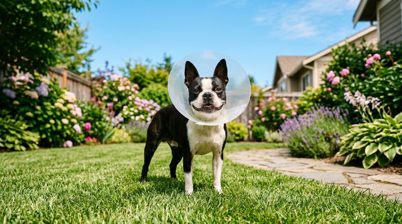
Maintaining your pet’s primary care with our independent, centrally located Tigard hospital ensures complete consistency of medical records and direct access to the exact surgical team that performed the procedure. Post-operative recovery requires close compliance, including wearing a protective electronic collar and returning for regular progress checks to ensure the dissolving sutures are healing correctly within the tissue pocket. Having a familiar, local medical team who knows your pet’s specific temperament makes these follow-up visits fast, low-stress, and highly reliable, allowing us to catch any early healing variations quickly.
Reclaiming Your Dog’s Optical Health and Visual Comfort
A prolapsed third eyelid gland will not slide back into its proper anatomical position permanently on its own, and delaying professional surgical repair only increases the risk of lifelong dry eye, chronic secondary infections, or painful corneal ulceration. Watching a bright pink mass grow in the corner of your dog’s eye can be deeply concerning, but choosing an independent, technologically advanced veterinary team ensures your pet receives modern, tissue-preserving pocket-repair surgery that protects their long-term vision and daily comfort. You do not have to leave your pet’s eye care to guesswork or wait out concerning tissue changes in silent worry.
The dedicated medical team at Nimbus Pet Hospital is fully prepared to provide the advanced surgical excellence, clear diagnostic imaging, and compassionate care your animal needs to heal completely. Our convenient location on SW Nimbus Ave provides local families with a reliable, high-tier partner for all aspects of preventative and surgical pet care. Take a definitive step to support your dog’s visual health before secondary bacterial contamination or self-inflicted scratching damages the eye permanently. Contact our friendly staff by calling or texting (971) 300-0613 right away, or stop by our hospital today to schedule a professional consultation and return clear, pain-free sight to your favorite companion.